ECG rhythms every RT should recognize
Normal sinus rhythm and its intervals, then sinus tachycardia and bradycardia, AF, flutter, PVCs, VT, VF, asystole, and PEA, with which ones are shockable.
- Sinus rate60–100/minBelow 60 bradycardia; above 100 tachycardia
- PR interval0.10–0.20 sLonger = first-degree AV block
- QRS0.07–0.10 s0.12 s or more is wide
- ShockableVF, pulseless VTAsystole and PEA are not
RTs are at the bedside when a rhythm changes, often because of hypoxemia, a drug, or a procedure. You don’t need to diagnose every strip. You do need to tell a patient who is fine from one who needs help or a defibrillator now.
The normal beat
The P wave is the atria firing. The QRS is the ventricles firing. The T wave is the ventricles resetting.
| Measure | Normal adult |
|---|---|
| Sinus rate | 60 to 100 per minute |
| PR interval (start of P to start of QRS) | 0.10 to 0.20 s |
| QRS width | 0.07 to 0.10 s |
A narrow QRS (under 0.12 s) means the beat started above the ventricles. A wide QRS (0.12 s or more) means it started in the ventricles, or travelled down a delayed pathway. Normal sinus rhythm has a P wave before every QRS, and the beats come at regular intervals.
Common rhythms
| Rhythm | What you see | Notes |
|---|---|---|
| Sinus tachycardia | Normal beats, rate over 100 | A response to hypoxemia, pain, fever, anxiety, or drugs such as bronchodilators. Treat the cause. |
| Sinus bradycardia | Normal beats, rate under 60 | Normal in athletes and sleep. In a sick patient, look for hypoxemia or a vagal response such as suctioning. |
| Atrial fibrillation (AF) | Irregularly irregular, no P waves, a quivering baseline | Rates above about 140 can drop cardiac output. |
| Atrial flutter | Regular “sawtooth” flutter waves, often a fixed block ratio | Can look regular or irregular depending on the block. |
| PVC | Early, wide QRS with no P wave, then a pause | Common and often harmless; frequent runs, or pairs, need attention. |
The dangerous ones
- Ventricular tachycardia (VT): three or more ventricular beats in a row at a rate of 120 or more (some use 100). The QRS is wide. Treat any wide-complex tachycardia as VT until proven otherwise. With a pulse the patient needs urgent help; without a pulse VT is shocked like VF.
- Ventricular fibrillation (VF): chaotic, ultrafast wiggles with no real QRS. The ventricles quiver and pump nothing. The patient is pulseless. Defibrillation is the treatment, and each minute of delay cuts the chance that the first shock works by about 10%.
- Asystole: no electrical activity, a flat line. Confirm in a second lead and check the leads and gain.
- Pulseless electrical activity (PEA): an organized rhythm on the monitor, but no pulse.
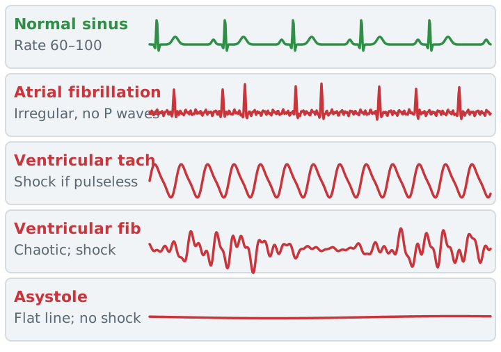
Which rhythms get a shock?
| Shockable | Not shockable |
|---|---|
| VF | Asystole |
| Pulseless VT | PEA |
Non-shockable rhythms get CPR and a search for the cause: the “Hs and Ts” (hypoxia, hypovolemia, acidosis, high or low potassium, hypothermia; toxins, tamponade, tension pneumothorax, thrombosis). Hypoxia and tension pneumothorax are in your lane. See CPR and resuscitation.
Artifact or real?
Patient movement, loose electrodes, and even tooth brushing can draw a tracing that looks like VT or VF. A monitor reading is not a patient. Look at the patient first: responsive, breathing, and a pulse mean it isn’t arrest. Check the other leads, because artifact often corrupts all but one lead, and the clean one may not be the one on the screen. If there is no pulse, start CPR.
Study example
During a bronchoscopy a monitor alarms for VT at a rate of 190. The patient is awake, talking, and has a good pulse and a normal SpO2 waveform. The strip is noisy in the lead on screen. You check the other leads and the electrode, and the other leads show normal beats. It was artifact from the patient’s movement. If he had been unresponsive and pulseless, you would start CPR and call for the defibrillator.
Related: CPR and resuscitation, hemodynamic monitoring, capnography.
Sources
- Merck Manual Professional Edition. Electrocardiography.
- Merck Manual Professional Edition. Overview of Arrhythmias.
- Merck Manual Professional Edition. Atrial Fibrillation.
- Merck Manual Professional Edition. Ventricular Tachycardia (VT).
- Merck Manual Professional Edition. Ventricular Fibrillation (VF).
- Merck Manual Professional Edition. Cardiac Arrest.
- Kacmarek RM, Stoller JK, Heuer AJ. Egan's Fundamentals of Respiratory Care. Elsevier. (Flutter waves, bronchodilator effects.)
- Drew BJ, et al. Insights into the problem of alarm fatigue with physiologic monitor devices. PLoS One. 2014.