Physical assessment: vitals, inspection, palpation, percussion
Normal adult vital signs and the bedside exam: work of breathing, cyanosis, clubbing, tracheal position, fremitus, percussion notes, and mental status.
- Respiratory rate12–18/minResting adult; some courses use 12–20
- Heart rate60–100/minResting adult
- Blood pressure90/60–120/80mmHg, resting adult
- Cyanosis needs≥ 5 g/dLUnsaturated hemoglobin; a late, unreliable sign
The bedside exam costs nothing and tells you how sick the patient is before any test comes back. Look first, then feel, then tap, then listen (breath sounds have their own topic). Compare one side with the other at every step.
Vital signs
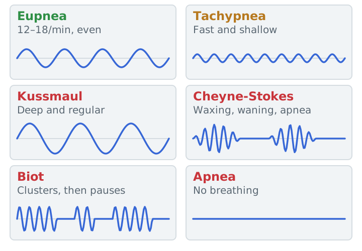
For a resting healthy adult, MedlinePlus gives a respiratory rate of 12 to 18 per minute, a heart rate of 60 to 100, and a blood pressure from 90/60 to 120/80 mmHg. Temperature runs about 36.5 to 37.3 °C. Count the respiratory rate for a full minute, because abnormal patterns make short counts misleading. Add SpO2 (normally 95 to 100%), but remember it says nothing about ventilation (pulse oximetry).
Inspection
- Work of breathing: a patient who can’t finish a sentence, sits upright, or braces the arms on the knees (tripod position) is working hard.
- Accessory muscles and retractions: use of the sternocleidomastoids, scalenes, and intercostals at rest means distress. Retractions show up between the ribs and above the clavicles when airflow is limited.
- Paradoxical breathing: the abdomen moves in during inspiration. It points to respiratory muscle fatigue or weakness. Treat it as a warning sign.
- Chest shape and pursed lips: a barrel chest (larger front-to-back diameter) goes with emphysema. Pursed-lip breathing slows expiration and creates a little back pressure that holds airways open.
- Clubbing: the nail angle flattens to more than 180° and the nail bed feels spongy. Causes include lung cancer, cystic fibrosis, and pulmonary fibrosis.
- Cyanosis is a blue tint of the lips and nail beds. It needs at least 5 g/dL of unsaturated hemoglobin, so by the time you see it, saturation is already well below normal. It is a late and unreliable sign: an anemic patient can be badly hypoxemic and look pink, and skin tone and room lighting change what you see.
- JVD and edema: with the patient at 45°, the jugular vein normally sits just above the clavicles. A neck vein that stays distended suggests right heart strain or failure. Swollen ankles with a raised jugular pressure point the same way.
Palpation
- Trachea: it should sit in the midline. A tension pneumothorax or large effusion pushes it away; atelectasis with volume loss pulls it toward the affected side.
- Chest expansion: put your hands on the lower ribs with the thumbs meeting at the midline and have the patient take a deep breath. Unequal movement suggests pneumothorax, pneumonia, rib fracture, or atelectasis.
- Tactile fremitus: feel the vibration while the patient says “ninety-nine.” It is increased over consolidation and decreased over a pleural effusion or pneumothorax.
Percussion
Tap the middle finger laid flat on the chest and compare matching spots.
| Note | Sounds like | Suggests |
|---|---|---|
| Resonant | Low, hollow | Normal air-filled lung |
| Hyperresonant | Louder, lower, drum-like | Hyperinflation (emphysema, asthma) or air in the pleural space |
| Dull | Short, thud-like | Fluid or solid tissue: effusion, consolidation, atelectasis, mass |
| Flat | Very short, no tone | Little or no air beneath: a large effusion, or over muscle and bone |
Obesity and a very muscular chest blunt the notes, so percussion is least reliable in those patients.
Level of consciousness
Describe what you see: alert, responds to voice, responds to pain only, or unresponsive. Restlessness and confusion can be early signs of hypoxemia. Growing drowsiness in a patient with COPD can mean CO2 retention, so look at the trend and confirm with an ABG.
Study example
A 68-year-old with COPD is sitting forward on his elbows, breathing at 28 with his lips pursed. You see sternocleidomastoid use and his abdomen moves in as his chest rises. The percussion note is hyperresonant on both sides, and the trachea is midline. This is increased work of breathing with early muscle fatigue, so call for help and get an ABG instead of trusting a normal-looking SpO2.
Related: breath sounds, chest X-ray basics, hemodynamic monitoring.
Sources
- MedlinePlus. Vital signs.
- Merck Manual Professional Edition. Evaluation of the Patient With Pulmonary Issues.
- Merck Manual Professional Edition. Pneumothorax.
- Toronto Metropolitan University. Posterior and Lateral Thorax: Percussion. In: Introduction to Health Assessment for the Nursing Professional.
- Poulin I. Respiratory System. In: Physical Assessment Essentials for Health Sciences. 2023 (CC BY 4.0).
- Kacmarek RM, Stoller JK, Heuer AJ. Egan's Fundamentals of Respiratory Care. Elsevier.