Lung volumes, capacities, and full PFTs
The four volumes and four capacities, which ones spirometry can't measure, obstructive versus restrictive patterns on full PFTs, and what DLCO adds.
- Tidal volume (VT)About 500 mLResting adult, about 7 mL/kg
- FRCAbout 40% of TLCRV + ERV; about 2.4 L of a 6 L TLC
- RestrictionTLC < LLNz-score below −1.645; needs lung volumes
- Needs more than spirometryRV, FRC, TLCHelium, N2 washout, or plethysmography
- Low DLCOEmphysema, ILDAlso pulmonary vascular disease, anemia
Spirometry measures what you can breathe in and out. It can’t measure the air that stays in the chest after the deepest exhalation. Full PFTs add that air, which is what separates trapped air and overinflation from true restriction.
Four volumes and four capacities
A volume is one slice of the breath. A capacity is two or more slices added together.
| Term | Meaning | Typical young adult male |
|---|---|---|
| Tidal volume (VT) | One resting breath | about 500 mL |
| Inspiratory reserve volume (IRV) | Extra air you can breathe in after a normal breath | about 3,100 mL |
| Expiratory reserve volume (ERV) | Extra air you can breathe out after a normal breath | about 1,200 mL |
| Residual volume (RV) | Air left after the hardest exhalation | about 1,200 mL |
| Inspiratory capacity (IC) | VT + IRV | about 3,600 mL |
| Functional residual capacity (FRC) | ERV + RV: what is left after a normal exhalation | about 2,400 mL |
| Vital capacity (VC) | IRV + VT + ERV: the most you can move | about 4,800 mL |
| Total lung capacity (TLC) | VC + RV: everything | about 6,000 mL |
Values vary with height, sex, age, and body size, so every result is compared with a predicted value for that person. FRC is about 40% of TLC. The same equations are on the formulas page.
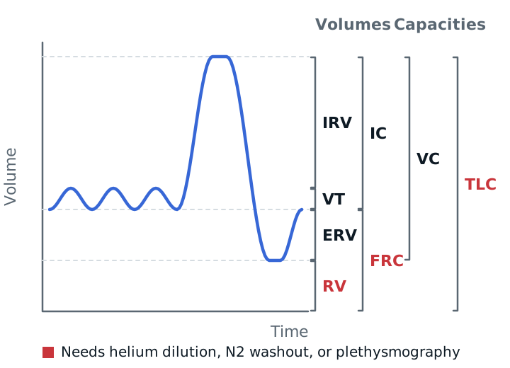
Measuring RV, FRC, and TLC
Spirometry stops at the air you can blow out, so it misses RV. Three methods measure FRC, and RV and TLC follow from it:
- Helium dilution: the patient breathes from a closed system containing a known amount of helium. The helium spreads out, and the drop in its concentration gives the lung volume.
- Nitrogen washout: the patient breathes 100% oxygen until exhaled nitrogen reaches zero. The total nitrogen collected gives the volume.
- Body plethysmography: the patient sits in a sealed box and pants against a closed mouthpiece. Boyle’s law turns the pressure changes into thoracic gas volume.
Helium and nitrogen only measure gas that connects to the airways, so trapped air can be missed and the volume underestimated. The box counts all of it, so it is more accurate in obstruction.
Obstructive versus restrictive
| Obstructive | Restrictive | |
|---|---|---|
| FEV1/FVC | Low | Normal or high |
| TLC | Normal or high | Low |
| RV | Normal or high | Low |
| RV/TLC ratio | High (air trapped) | Normal or high |
| FRC | Often high (hyperinflation) | Low |
Restriction means a TLC below the lower limit of normal (about the 5th percentile, z below −1.645). A low FVC with a normal ratio only suggests restriction; the TLC confirms it. Overinflation shows as a raised TLC, FRC, and RV.
DLCO
The diffusing capacity for carbon monoxide shows how well gas crosses from the alveoli into the blood. The patient inhales a small dose of CO, holds the breath, and exhales. DLCO depends on the surface area, the thickness of the barrier, and the amount of blood in the capillaries. It is read with alveolar volume and hemoglobin.
- Low: emphysema, pulmonary fibrosis and other interstitial disease, pulmonary vascular disease (pulmonary hypertension, emboli), and anemia.
- High: polycythemia, alveolar hemorrhage, and sometimes asthma.
An obstructive pattern with a low DLCO points toward emphysema. A restrictive pattern with a low DLCO points toward interstitial disease. An isolated low DLCO with normal volumes can be the earliest sign of interstitial or pulmonary vascular disease, or severe anemia.
Study example
A patient’s FEV1/FVC is 0.84, FVC is low, TLC is well below the lower limit of normal, and DLCO is low. Normal ratio plus a low TLC confirms restriction. The low DLCO says the problem reaches the gas-exchange surface, which fits interstitial lung disease more than a chest wall problem.
Related: spirometry basics, COPD, asthma.
Sources
- Merck Manual Professional Edition. Airflow, Lung Volumes, and Flow-Volume Loop.
- Merck Manual Professional Edition. Measurement of Gas Exchange.
- American Thoracic Society. PFT Interpretation: Resident Boot Camp teaching slides. 2024.
- Kacmarek RM, Stoller JK, Heuer AJ. Egan's Fundamentals of Respiratory Care. Elsevier. (Typical adult values.)