Chest X-ray basics for the RT
PA versus AP films, a systematic read, tissue densities, and key findings: hyperinflation, consolidation, atelectasis, pneumothorax, effusion, edema, and tubes.
- Hyperinflation> 10 posterior ribsAdequate inspiration shows about 8–10
- Heart size (PA film)< ½ chest widthAP films magnify it
- ETT tipAbout 5 cm above carinaHead neutral; range about 2–6 cm
- Effusion bluntingAbout 175 mLLateral angle on an upright film
You will look at chest films daily: after intubation, after a line goes in, and when a patient gets worse. You are not expected to report them, but you must be able to spot a tube in the wrong place, a collapsed lung, or air and fluid in the pleural space, and tie them to what you hear and see at the bedside.
PA versus AP
A PA film is taken upright in the radiology suite with the x-ray source about 6 feet away and the patient’s chest against the detector. An AP film is usually a portable bedside film with the source about 3 feet away. The shorter distance and the heart’s position farther from the detector magnify the heart and mediastinum, so an AP film can suggest cardiomegaly or a wide mediastinum that isn’t there. Portable films are often supine and taken with a poor breath in, which can also mimic edema.
Lateral views add depth. A lateral decubitus view (lying on the side) shows whether effusion fluid is free and moves with gravity, and an end-expiratory film can bring out a small pneumothorax.
Densities
| Looks | Tissue |
|---|---|
| Black | Air |
| Dark gray | Fat |
| Bright gray | Water: soft tissue, blood, heart, fluid |
| White | Bone, metal (tubes, wires) |
Two structures of the same density touching each other blur together. Lung that fills with fluid or pus turns water density and loses its edge against the heart or diaphragm. This is the silhouette sign. A lost right heart border points to the right middle lobe; a lost left heart border points to the lingula.
A systematic read
- Check the film. Patient, date, side marker, PA or AP, upright or supine.
- Inspiration. The diaphragm should sit at about the 8th to 10th posterior rib.
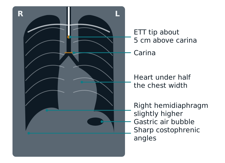
- Airway. Trachea midline? Where are the tube tip and the carina?
- Lungs. Compare the two sides zone by zone. Look for white areas, black areas, and vessels running out to the edge.
- Pleura and diaphragm. Sharp costophrenic angles, a normal dome (the right is slightly higher).
- Heart and mediastinum. On a PA film the heart is under half the chest width and the mediastinum under about 8 cm.
- Bones and lines. Ribs, then every tube and line.
Findings you must know
| Finding | What you see |
|---|---|
| Hyperinflation (COPD, asthma) | More than 10 posterior ribs visible; flattened diaphragms; dark, wide lungs. |
| Consolidation | A white patch of alveolar filling. Air bronchograms (dark branching airways inside it) prove the alveoli are filled, but not that the cause is infection. A silhouette sign if it touches the heart or diaphragm. |
| Atelectasis | Volume loss: trachea, heart, and fissures pulled toward the collapsed side, a raised hemidiaphragm, crowded vessels, and a denser lobe. |
| Pneumothorax | No lung markings past a thin visceral pleural line. Supine, air collects in front, so look for a deep sulcus (a deeper, darker costophrenic angle). Tension pushes the mediastinum away and flattens the hemidiaphragm. |
| Pleural effusion | Uniform density that blunts the costophrenic angle; on an upright film fluid climbs the chest wall, forming a meniscus. A large effusion pushes the mediastinum away. |
| Pulmonary edema | Upper-zone vessels enlarge (cephalization), Kerley B lines and cuffing around the airways, then patchy, bat-wing alveolar fluid, often with a big heart and effusions. |
About 75 mL of fluid blunts the back angle on an upright film and about 175 mL blunts the side angle. Collapse pulls the mediastinum toward the lesion and an effusion does not, so shift direction separates them.
Tubes and lines
An endotracheal tube tip should lie about 5 cm above the carina with the head neutral; the accepted range is roughly 2 to 6 cm. Flexing the neck moves the tube deeper, and extension pulls it out. A tube that is too deep enters the right mainstem bronchus and the left lung collapses, with the heart and trachea pulled toward it.
A film shows how deep the tube is, not whether it is in the trachea or esophagus. Both lie in the midline behind each other. Confirm placement with waveform capnography (endotracheal intubation).
Study example
After intubation, a portable film shows the tube passing below the carina into the right main bronchus, a dense left lung, and the trachea shifted left. Breath sounds are quiet on the left. The tube is in the right mainstem and the unventilated left lung has collapsed. Pull the tube back a few centimeters, check breath sounds, and repeat the film.
Related: endotracheal intubation, physical assessment, breath sounds, ARDS.
Sources
- Broder J. Imaging the Chest: The Chest Radiograph. In: Diagnostic Imaging for the Emergency Physician. Elsevier; 2011:185-296.
- Jain SN, Modi T, Aswani Y, Varma RU. Chest radiography in adult critical care unit: a pictorial review. Indian J Radiol Imaging. 2019;29(4):418-425.
- Merck Manual Professional Edition. Chest Imaging.
- Merck Manual Professional Edition. Pleural Effusion.
- Merck Manual Professional Edition. Pneumothorax.