Study notesTherapeutic procedures
Chest tubes and the three-chamber drainage system
Why chest tubes are placed, what each drainage chamber does, how to read tidaling and bubbling, and what to do if the tube disconnects or the unit tips.
- Water seal depth2 cmOne-way valve; keep at the fill line
- TidalingNormalFluid rises and falls with breathing
- Bubbling in water sealAir leakCheck patient, tube, and connections
- Typical suction−20 cm H2OCommon initial set level; ordered by the physician
- Unit positionBelow chestAlways, including during transport
A chest tube (chest drain) is a flexible tube placed through the chest wall into the pleural space to remove air or fluid so the lung can re-expand. It connects to a drainage unit that lets air and fluid out but does not let air back in. RTs see these patients on the ventilator, on the floor after surgery, and in transport, so you need to read the unit at a glance.
Why a chest tube is placed
Common indications are pneumothorax, hemothorax, pleural effusion, and empyema (infected pleural fluid). Others are chylothorax and drainage after chest surgery or trauma. See pneumothorax and pleural effusion.
The three chambers
Most plastic units have three connected chambers.
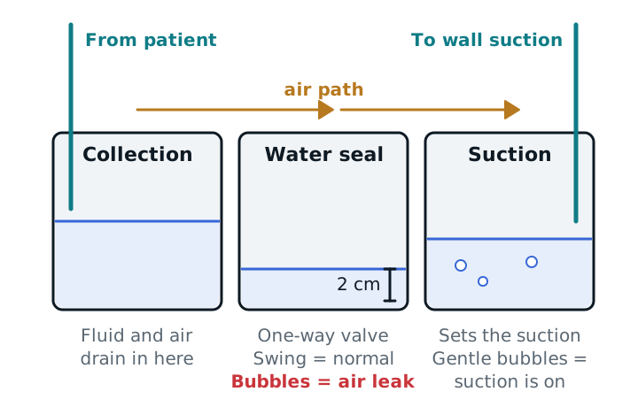
- Collection chamber. Fluid and air from the pleural space drain here. Mark the level and note the color (serous, bloody, cloudy) and amount. A sudden bright-red increase or a change to cloudy fluid needs a call to the team.
- Water seal chamber. A column of water, about 2 cm, acts as a one-way valve. Air can leave the chest but cannot be pulled back in on inspiration.
- Suction control chamber. It sets how much suction is applied. A wet system uses a water column whose height sets the pressure (a water column tops out near −25 cm H2O), so gentle, continuous bubbling means suction is on. A dry system uses a dial or regulator and can go as high as −40 cm H2O; an indicator shows that suction is working. Suction is not always needed. It can prolong an air leak in some patients, so many tubes sit on water seal (gravity drainage).
Wall suction does not set the pressure by itself: the chamber or dial does. The usual starting level is −20 cm H2O, but the physician orders it.
Reading the water seal
- Tidaling (swing). The water level in the water seal chamber rises and falls with breathing. It means the tube is open and connected to the pleural space. Seeing it is normal.
- No tidaling. The lung may have fully re-expanded and plugged the tube holes, or the tube is kinked, clotted, or out of the pleural space. Check the tubing for kinks and tell the physician if you find none.
- Intermittent bubbling when the patient exhales or coughs means an air leak from the lung. Many leaks seal on their own. Continuous bubbling means a large leak, or air entering through a loose connection or a tube that has slipped so a drainage hole is outside the skin. Check the connections and the insertion site.
Problems and what to do
- Tube disconnects from the unit. If the drain was bubbling up to the moment it came apart, reconnect right away. Otherwise some protocols clamp briefly at the patient end, clean the ends, attach sterile tubing, and unclamp. Other protocols place the open end in sterile water to recreate a water seal. Follow your facility’s protocol, then assess the patient, check vital signs, and notify the physician (a chest X-ray may be ordered).
- Unit tips over. Stand it upright right away and confirm the water seal is at the fill line. Add sterile water if needed. If the patient is well and the unit drains, no more action is needed; watch the patient.
- Tube pulls partly or fully out. Cover the site with an occlusive dressing, call for help, and assess for subcutaneous emphysema and distress.
- Large effusion. Large pleural effusions are drained in a controlled way (the BTS standard is to stop at 1,000 mL) to lower the risk of re-expansion pulmonary edema.
- Moving the patient. Keep the unit below chest level, never lift it above the chest, and keep the tubing free of kinks. Some protocols disconnect suction and leave the drain open to air during transport.
Study example
You are ventilating a trauma patient with a left chest tube on water seal. At the start of the shift the water seal rises and falls with each breath and nothing bubbles. After a coughing fit you see bubbles in the water seal on each exhalation, and the collection chamber is unchanged. That fits a small air leak. You check that every connection is tight and the unit is below the chest, watch the patient’s SpO2, and report the finding. You do not clamp the tube.
See pneumothorax and pleural effusion and lung expansion therapy.
Sources
- Porcel JM. Chest Tube Drainage of the Pleural Space: A Concise Review for Pulmonologists. Tuberc Respir Dis. 2018;81(2):106-115.
- British Thoracic Society. Online Appendix 8: How to set up a chest drain bottle/underwater seal drain (pleural procedures).
- Royal Children's Hospital Melbourne. Clinical Practice Guideline: Chest drain management.
- StatPearls. Care of a Chest Tube. NCBI Bookshelf.