Pneumothorax and pleural effusion
Recognize a pneumothorax, spot a tension pneumothorax on a ventilator, and tell transudate from exudate with Light's criteria.
- Needle decompression (adult)4th–5th ICSSide of chest, anterior-to-mid axillary line (ATLS)
- Exudate: protein ratio> 0.5Pleural fluid / serum protein (Light's)
- Exudate: LDH ratio> 0.6Or pleural LDH above two-thirds of normal serum
The pleural space is the thin gap between the lung and the chest wall. Air in it is a pneumothorax; fluid in it is a pleural effusion. Both let the lung down, both cause shortness of breath, and both show up in an RT’s day, especially on a ventilator.
Pneumothorax
Types:
- Primary spontaneous: no obvious lung disease; often small apical blebs. Smoking and a tall, thin build raise the risk.
- Secondary spontaneous: a complication of existing lung disease, such as COPD.
- Traumatic or iatrogenic: from a chest injury, a central line, a biopsy, or positive pressure ventilation.
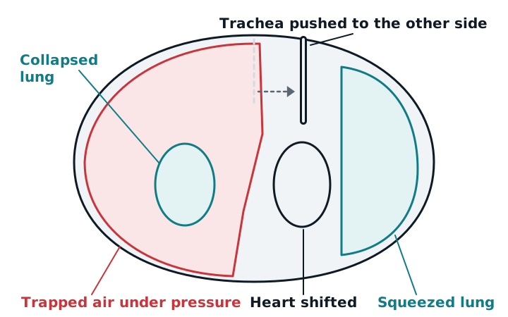
- Tension: air enters the pleural space with each breath and can’t leave. Pressure builds, pushes the mediastinum toward the other side, and squeezes the other lung and the great veins.
Signs
On the affected side you find decreased or absent breath sounds and a hyperresonant percussion note, which sounds tympanitic when the pneumothorax is under tension. ATLS lists acute respiratory distress, subcutaneous emphysema, absent breath sounds, hyperresonance, and a tracheal shift as the signs that call for immediate decompression. In a ventilated patient, the first sign is often sudden: peak airway pressure climbs, minute ventilation drops, SpO2 falls, and blood pressure drops. Treat that combination as a tension pneumothorax until proven otherwise, and rule out a circuit or tube problem while help is on the way. See initial ventilator settings for what normal pressures look like.
Imaging
On a chest X-ray, look for a visceral pleural line: a thin line separated from the chest wall by a lucent space with no lung markings. Skin folds can mimic it. On a supine film, air collects in front rather than at the apex, and a deep sulcus sign (a deep, dark costophrenic angle) may be the only clue. Ultrasound shows absent lung sliding.
Treatment
A tension pneumothorax is a clinical diagnosis; don’t wait for an X-ray. The immediate fix is needle decompression. The current ATLS site in adults is the 4th or 5th intercostal space on the side of the chest, in the anterior-to-mid axillary line, with a 5 to 8 cm catheter. It replaced the older 2nd intercostal space, midclavicular line site in 2018, which some other guidelines still use. A chest tube follows. For a stable patient with a primary spontaneous pneumothorax, a French guideline advises watching a small one and using needle aspiration or a chest tube for a large one.
Pleural effusion
An effusion is extra fluid in the pleural space. On exam you find dullness or flatness to percussion, decreased fremitus, and absent or diminished breath sounds over the fluid. A large effusion pushes the trachea and heart away from that side. The upright chest X-ray shows blunting of the costophrenic angle, a rounded, white angle where the fluid sits.
Thoracentesis (needle drainage) is both treatment and a test, and ultrasound is used to guide it. The fluid is sorted into transudate or exudate:
- Transudate: from pressure imbalance, as in heart failure.
- Exudate: from inflammation, infection, or cancer in the pleura.
By Light’s criteria, the fluid is an exudate if any of these is true: pleural/serum protein above 0.5, pleural/serum LDH above 0.6, or pleural LDH above two-thirds of the upper limit of normal serum LDH. The criteria sometimes call a transudate an exudate (about 16% of transudates in one study), so the result is read with the clinical picture.
Study example
A patient on volume control has a rising peak pressure alarm, SpO2 falling from 95% to 82%, and blood pressure of 80/50. Breath sounds are absent on the left and the trachea has shifted to the right. You disconnect and hand-bag to rule out a circuit problem, tell the team, and the physician decompresses the left chest.
Related: asthma, COPD, heart failure, pulmonary embolism.
Sources
- Spontaneous Pneumothorax: A Review of Underlying Etiologies and Diagnostic Imaging Modalities. Tomography. 2025.
- Murray and Nadel's Textbook of Respiratory Medicine: History and Physical Examination (Table 16-3).
- Paramedic Understanding of Tension Pneumothorax and Needle Thoracostomy (NT) Site Selection. Cureus. 2022.
- Emergent needle thoracostomy in prehospital trauma patients: a review of procedural execution through computed tomography. Trauma Surg Acute Care Open. 2021.
- Pleural effusion in critically ill patients and intensive care setting. World J Clin Cases. 2023.
- SPLF/SMFU/SRLF/SFAR/SFCTCV Guidelines for the management of patients with primary spontaneous pneumothorax. Ann Intensive Care. 2023.