Hypoxemia: causes, A–a gradient, and oxygen delivery
The five causes of hypoxemia, how the A–a gradient and oxygen response sort them, hypoxia types, the P/F ratio, and oxygen content and delivery.
- HypoxemiaPaO2 < 60 mmHgOr SaO2 < 90%
- Normal A–a gradient< (age/4) + 4Usually under 20 mmHg on room air
- ShuntRefractory to O2Raising FiO2 adds little
- P/F ratioPaO2 ÷ FiO2FiO2 as a fraction; ARDS bands 300, 200, 100
- CaO21.34 × Hb × SaO2 + 0.003 × PaO2mL O2/dL blood
Hypoxemia is a low oxygen level in arterial blood: generally a PaO2 below 60 mmHg or an SaO2 below 90%. Hypoxia is low oxygen at the tissues, and it can happen with a normal PaO2. Courses grade hypoxemia as mild, moderate, or severe with different cutoffs, so use your instructor’s bands.
Five causes
- Low inspired oxygen, mostly altitude.
- Hypoventilation: too little alveolar ventilation. PaCO2 rises as PAO2 and PaO2 fall.
- V/Q mismatch: ventilation and blood flow don’t line up. This is the usual cause in COPD and asthma.
- Shunt: blood passes through the lungs without touching ventilated alveoli, such as through collapsed or fluid-filled air spaces (atelectasis, pneumonia, edema, ARDS). Shunt is the extreme end of low V/Q.
- Diffusion limitation: gas can’t cross a thickened membrane in time. Rarely the sole cause; it shows up during exercise (blood moves through faster) or at altitude.
The A–a gradient sorts them
The A–a gradient is calculated PAO2 minus measured PaO2. Calculate PAO2 as FiO2 × (PB − 47) − PaCO2/0.8. Normal is less than about (age/4) + 4, and usually under 20 mmHg on room air. It rises with age and with higher FiO2. For PaO2 70 and PaCO2 40 on room air at sea level, PAO2 is about 100, and the gradient is 30: widened.
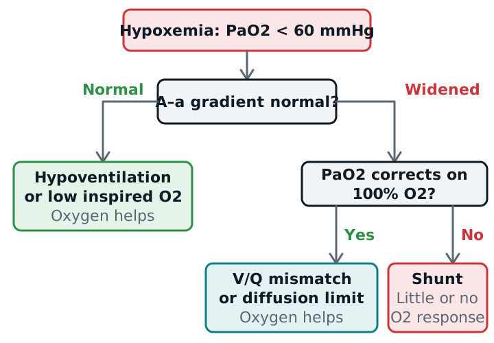
- Normal gradient: low inspired O2 and hypoventilation. The lungs are fine; the problem is how much gas gets in. Check the PaCO2: a high value means hypoventilation.
- Widened gradient: V/Q mismatch, shunt, and diffusion limitation. The lung itself is the problem.
Does oxygen fix it?
Low inspired O2, hypoventilation, V/Q mismatch, and diffusion limitation all respond to supplemental oxygen. A true shunt does not: the shunted blood never meets the extra oxygen, and the blood that does pass through healthy alveoli is already nearly full. Shunt needs the alveoli reopened (PEEP, treating the cause), not just more FiO2.
Four kinds of hypoxia
| Type | Problem | Example | PaO2 |
|---|---|---|---|
| Hypoxemic | Too little O2 in arterial blood | Pneumonia, COPD | Low |
| Anemic | Too little or abnormal hemoglobin | Anemia, carbon monoxide | Normal |
| Circulatory (stagnant) | Too little blood flow | Shock, heart failure | Normal |
| Histotoxic | Cells can’t use the oxygen | Cyanide poisoning | Normal |
Only the first is helped much by a higher FiO2. In the others, correct the real problem: hemoglobin, flow, or the poison.
P/F ratio
P/F = PaO2 ÷ FiO2, with FiO2 as a fraction (40% = 0.40). A healthy adult on room air is about 100 ÷ 0.21, roughly 475. The Berlin ARDS bands are 300, 200, and 100, with PEEP of at least 5 (ARDS). The ratio lets you compare patients on different FiO2 values, but it changes with PEEP and the amount of shunt.
Oxygen content and delivery
CaO2 = (1.34 × Hb × SaO2) + (0.003 × PaO2) in mL O2/dL. Hemoglobin carries almost all of it; the dissolved part is tiny. Some references use 1.31 or 1.36 for the 1.34, which changes the answer slightly. DO2 = CO × CaO2 × 10 mL/min, and a resting adult’s is about 1,000 mL/min. See the formulas page.
With Hb 15, SaO2 0.98, PaO2 100: CaO2 = 19.7 + 0.3 = 20 mL/dL. With a cardiac output of 5 L/min, DO2 is about 1,000 mL/min. If Hb falls to 7.5 and nothing else changes, DO2 halves, even though PaO2 and SpO2 are unchanged.
Study example
A patient on room air has PaO2 52 and PaCO2 38. PAO2 is 0.21 × 713 − 38/0.8 = about 102, so the A–a gradient is about 50: widened, so the lung is involved and the PaCO2 rules out hypoventilation. Placed on 100% oxygen, the PaO2 rises only to 70. That is a poor response: shunt, as in pneumonia or ARDS. The next step is to look for lung that can be recruited, not just to turn up the FiO2.
Related: ABG interpretation, pulse oximetry, oxygen therapy principles, ARDS.
Sources
- Merck Manual Professional Edition. Measurement of Gas Exchange.
- Ghan T. Oxygen Physiology. OpenAnesthesia. 2024.
- Merck Manual Professional Edition. Evaluation of the Patient With Pulmonary Issues.
- Kacmarek RM, Stoller JK, Heuer AJ. Egan's Fundamentals of Respiratory Care. Elsevier.