Study notesTherapeutic procedures
Lung expansion therapy: incentive spirometry, PEP, CPAP, IPPB
How to prevent and treat atelectasis: incentive spirometry technique, what the AARC says about it, when to move to CPAP, PEP, or IPPB, and signs of atelectasis.
- IS breath-hold≥ 5 secondsSustained maximal inspiration
- IS dosing example10 breaths / 1–2 hAwake; AARC found no proven frequency
- PEP pressure10–20 cm H2ODuring exhalation
- CPAP pressure5–20 cm H2OThroughout the breath
- IPPB goal≥ 1/3 of predicted ICMinimum delivered tidal volume
Lung expansion therapy uses techniques that raise lung volume to prevent or reverse atelectasis (collapsed alveoli). It matters most after abdominal or chest surgery, with prolonged bed rest, or with a weak cough, because shallow breathing and poor secretion clearance let alveoli close.
Signs of atelectasis
Look for a rising respiratory rate, tachycardia, low-grade fever, diminished breath sounds over the affected area, a falling SpO2 or rising FiO2 need, and a chest X-ray showing volume loss. When therapy works, the same findings reverse: the rate and pulse fall, breath sounds return, and oxygenation improves (AARC incentive spirometry guideline).
Incentive spirometry
Incentive spirometry (IS), also called sustained maximal inspiration, copies a natural sigh or yawn. A slow, deep breath lowers pleural pressure and expands the lungs. The device gives the patient visual feedback toward a target.
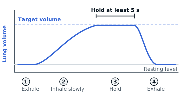
- Sit upright if you can. Hold the device upright.
- Breathe out normally.
- Seal your lips on the mouthpiece.
- Breathe in slowly and deeply to raise the indicator toward the target. Slow is the point: a fast sniff raises the ball but moves little volume.
- At the top, remove the mouthpiece and hold your breath for at least 5 seconds.
- Breathe out normally, rest, and repeat.
The AARC suggests a volume-oriented device, which usually adds less work of breathing and gives larger inspiratory volumes than a flow-oriented one. Evidence for a specific frequency is lacking. Trials have used 10 breaths every 1–2 hours while awake. Teach the technique, watch a return demonstration, and then leave the device within reach.
Who can use it: the patient must be awake, cooperative, and able to take a deep breath. IS is contraindicated when the patient cannot breathe deeply because of pain, diaphragm dysfunction, or opioid sedation, or cannot follow instructions (confused, heavily sedated, very young). Treat pain first. Possible hazards are hyperventilation, fatigue, pain, and hypoxemia if the patient takes off prescribed oxygen to use the device.
What the AARC position says
The 2011 AARC guideline is blunt: IS alone is not recommended for routine use to prevent postoperative pulmonary complications. It recommends IS together with deep-breathing technique, directed coughing, early mobilization, and good pain control. Deep-breathing exercises are suggested to work as well as IS. Routine IS is not recommended to prevent atelectasis after upper-abdominal or CABG surgery. Getting the patient up and walking helps because upright posture and activity raise lung volume.
When IS is not enough
| Therapy | How it works | Use when |
|---|---|---|
| CPAP | One constant pressure, 5–20 cm H2O, through the whole breath | Atelectasis that does not respond to deep breathing; the patient breathes spontaneously |
| PEP | Patient exhales against a resistor, usually 10–20 cm H2O | Atelectasis, and to help mobilize secretions (add coughing) |
| IPPB | Machine-delivered positive-pressure breath for lung expansion | Significant atelectasis when other methods fail, or the patient cannot cooperate or deep breathe |
The 1993 PAP guideline lists air trapping, retained secretions, and atelectasis as indications. It lists untreated pneumothorax, hemodynamic instability, recent facial or skull surgery, active hemoptysis, nausea, and elevated ICP (above 20 mmHg) among the conditions to evaluate before using a mask. CPAP and PEP need the patient to breathe on their own.
The IPPB guideline treats IPPB as not the first choice when cheaper therapy can do the job. For lung expansion, aim for a delivered tidal volume of at least one-third of predicted inspiratory capacity. Hazards include barotrauma, gastric distention, hyperventilation, and increased work of breathing. An untreated tension pneumothorax is an absolute contraindication.
Study example
A 68-year-old man on postoperative day 1 after upper-abdominal surgery has a respiratory rate of 26, SpO2 91% on 2 L/min, and diminished breath sounds at the left base. He tries IS but only reaches a third of his preoperative target because incision pain stops him. You ask the team about pain control, get him sitting up and walking, and coach slow breaths with a 5-second hold. If he cannot cooperate even after analgesia, you discuss CPAP or IPPB with the physician.
See chest physiotherapy devices, noninvasive ventilation, and chest tubes.
Sources
- Restrepo RD, Wettstein R, Wittnebel L, Tracy M. AARC Clinical Practice Guideline: Incentive Spirometry, 2011. Respir Care. 2011;56(10):1600-1604.
- AARC Clinical Practice Guideline: Intermittent Positive Pressure Breathing, 2003 revision and update. Respir Care. 2003;48(5):540-546.
- AARC Clinical Practice Guideline: Use of Positive Airway Pressure Adjuncts to Bronchial Hygiene Therapy. Respir Care. 1993;38(5):516-521.
- Wilkins RL. Lung expansion therapy. In: Kacmarek RM, Stoller JK, Heuer AJ. Egan's Fundamentals of Respiratory Care. Elsevier.