Obstructive sleep apnea: AHI, scoring, and PAP
How polysomnography scores apneas and hypopneas, the AHI severity bands, obstructive versus central events, and CPAP, APAP, and bilevel.
- Apnea≥ 90% flow dropFor 10 seconds or longer
- Hypopnea (AASM 2012)≥ 30% flow drop10 s or longer, with ≥ 3% desaturation or an arousal
- AHI: mild / mod / severe5–14 / 15–29 / ≥ 30Events per hour of sleep
- Obesity hypoventilationBMI ≥ 30, PaCO2 ≥ 45Awake, after excluding other causes
Obstructive sleep apnea (OSA) is repeated collapse of the upper airway during sleep. The patient keeps trying to breathe, but the airway is blocked, so airflow stops or drops, oxygen falls, and the brain wakes briefly to reopen the airway. RTs meet OSA in the sleep lab, in the hospital, and above all when fitting masks and teaching patients to use PAP.
How it is measured
An overnight polysomnography (PSG) records airflow, chest and abdominal effort, oxygen saturation, and sleep stages. Technologists score each breathing event using AASM rules:
- Apnea: airflow falls by 90% or more from baseline for at least 10 seconds.
- Hypopnea: airflow falls by 30% or more for at least 10 seconds, with a drop in SpO2 of at least 3% or an arousal from sleep (the AASM 2012 rule). Earlier AASM versions used other cutoffs, such as a 4% desaturation, so the scoring rule changes the AHI.
The apnea-hypopnea index (AHI) is the number of events per hour of sleep. An AHI of 5 or more is the usual cutoff for OSA, and severity follows these bands:
| AHI (events/hour) | Category |
|---|---|
| Below 5 | Normal |
| 5 to 14 | Mild |
| 15 to 29 | Moderate |
| 30 or more | Severe |
Obstructive, central, or mixed
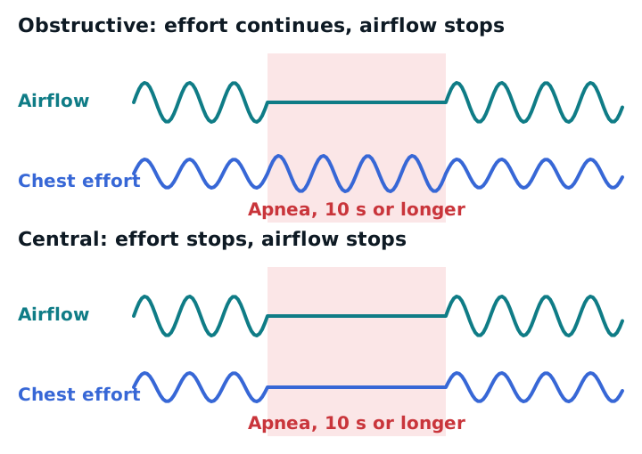
The tracing tells you which one it is by looking at effort:
- Obstructive: airflow stops, but the chest and abdomen keep moving. The patient is trying to breathe against a blocked airway.
- Central: airflow stops and effort stops too. The brain isn’t sending the drive to breathe. Causes include heart failure, neurologic disease, opioids, and high altitude.
- Mixed: an event that starts without effort and ends with effort.
Central sleep apnea is defined by central events making up more than half of all events, with at least 5 events an hour. It is treated by addressing the cause, and PAP, adaptive servo-ventilation, or other therapy may be used.
Treatment with PAP
CPAP holds the airway open with a fixed pressure. APAP (auto-titrating) adjusts the pressure within a range as the airway changes through the night. Bilevel PAP gives a higher pressure on inhalation than on exhalation.
The AASM guideline for adults with OSA says:
- Use PAP for OSA with excessive sleepiness (strong recommendation).
- Either CPAP or APAP is appropriate for ongoing treatment (strong); for routine use they are suggested over bilevel (conditional).
- Bilevel is a clinical-judgment choice, for example when pressure needs exceed what CPAP or APAP can deliver.
- For a patient without significant comorbidities, start with home APAP or an in-lab titration.
- Give education when PAP starts, and use troubleshooting and follow-up, including remote monitoring of use data, in the first weeks.
Your day-to-day work is mask fit, comfort, humidification, teaching, and checking that the patient actually uses the device.
Obesity hypoventilation syndrome
Obesity hypoventilation syndrome (OHS) is a BMI of 30 or more with awake hypercapnia (PaCO2 of 45 mmHg or more) after other causes are ruled out. About 90% of patients with OHS also have OSA, and around 70% have an AHI of 30 or higher. Treatment includes weight loss and PAP: CPAP if OSA is the main problem, and NIV when hypoventilation persists.
Study example
A sleep study shows 160 apneas and hypopneas over 5 hours of sleep. AHI = 160 ÷ 5 = 32 events per hour, which is severe. Most events show chest effort during the airflow pause, so this is obstructive. The sleep physician orders APAP titration. At the first follow-up, you check the mask seal and the data downloaded from the device.
Related: noninvasive ventilation, capnography, COPD.
Sources
- Kapur VK, et al. Treatment of Adult Obstructive Sleep Apnea with Positive Airway Pressure: An American Academy of Sleep Medicine Clinical Practice Guideline. J Clin Sleep Med. 2019.
- Impact of Hypopnoea Classifications on the Assessment of Sleep Disordered Breathing in Heart Failure. Card Fail Rev. 2025.
- Sleep-Disordered Breathing: Key Pathophysiological Mechanisms and Their Clinical Implications. Life (Basel). 2026.
- A systematic review of obstructive sleep apnea in Indigenous peoples worldwide. Sleep Adv. 2026 (reports the AHI severity bands used in the studies it reviews).