Study notesMechanical ventilation
Ventilator graphics: scalars and loops
Read pressure, flow, and volume waveforms to spot auto-PEEP, leaks, resistance versus compliance changes, asynchrony, and overdistension.
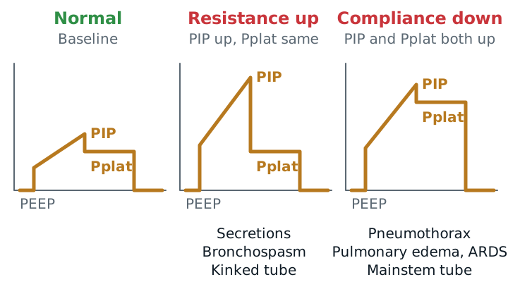
- Resistance problemPIP up, Pplat sameGap between PIP and Pplat widens
- Compliance problemPIP and Pplat upGap stays about the same
- Auto-PEEP signFlow not back to 0Before the next breath starts
A ventilator screen draws the patient’s breathing as curves. Read them in a fixed order and most problems show up before the alarm does.
The three scalars
A scalar plots one variable against time.
- Pressure-time: baseline is PEEP. The top of the curve is the peak inspiratory pressure (PIP). In volume control (VC), pressure rises steadily because flow is constant. In pressure control (PC), the curve is square.
- Flow-time: inspiration is above zero, expiration below. In VC the inspiratory block is square. In PC it starts high and decelerates. Exhalation is passive, so the expiratory curve depends on resistance and compliance.
- Volume-time: rises with inspiration and falls with expiration. Inspired and expired volume should match.
PIP versus plateau: resistance or compliance?
Hold the end of inspiration for 0.5 seconds in VC and pressure drops from PIP to the plateau pressure (Pplat). PIP reflects resistance plus compliance. Pplat reflects mainly the stiffness of the lungs and chest wall.
| Finding | Meaning | Common causes |
|---|---|---|
| PIP up, Pplat unchanged | Resistance increased | Secretions, bronchospasm, kinked or bitten tube |
| PIP and Pplat both up | Compliance decreased | Pneumothorax, pulmonary edema, ARDS, right mainstem tube, abdominal distension |
A sawtooth pattern on the flow or volume waveform, along with coarse breath sounds, is a sign of secretions and an indication to suction.
Auto-PEEP
If the next breath starts before exhalation finishes, gas is trapped and the alveolar pressure at end-expiration is above the set PEEP. That is auto-PEEP (intrinsic PEEP). It is common in COPD and asthma, and with fast rates or long inspiratory times.
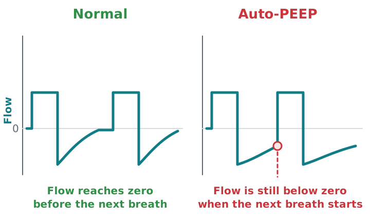
The flow curve shows that it is there; an expiratory hold measures it. Consequences are overdistension, lower blood pressure from less venous return, and missed triggers (the patient must first overcome the trapped pressure). Fix it by lengthening expiratory time: lower the rate, shorten inspiratory time, raise flow, treat bronchospasm, and sedate or adjust the mode if the patient is over-breathing the set rate.
Leaks
Exhaled volume that is less than inspired volume means gas is leaking: a cuff leak, circuit disconnect, chest tube air leak, or bronchopleural fistula. On the volume scalar the curve fails to return to zero. A trapped-gas problem can look similar, so check the flow-time scalar too.
Asynchrony
Asynchrony means the patient and the ventilator disagree about timing. Look for:
- Ineffective efforts: a small dip in pressure or flow with no breath. Auto-PEEP or low sensitivity can be the cause.
- Auto-triggering: breaths with no patient effort. Check for leak, circuit condensate, or cardiac oscillations, and re-set the trigger.
- Double triggering: two breaths back to back. The inspiratory time is too short or the volume too low for what the patient wants.
- Flow starvation: the patient pulls harder than the set flow supplies. In VC, raise the flow or switch to a mode where flow varies with effort.
- Premature or delayed cycling: the breath ends too early or too late in pressure support.
Loops
A pressure-volume loop that flattens at the top into a “beak” suggests overdistension: more pressure gives little more volume. A flow-volume loop whose expiratory limb is scooped out with a low peak flow suggests airflow obstruction, such as bronchospasm.
Study example
A COPD patient in VC at 18/min has a PIP of 32 and a Pplat of 20 cm H2O. The flow-time scalar shows expiratory flow still below zero when the next breath begins. The wide PIP-Pplat gap points to airway resistance, and the unfinished expiration points to air trapping. You lower the rate to 12/min and shorten inspiratory time, then recheck the flow curve and the blood pressure.
See ventilator modes, alarms and troubleshooting, initial settings, and COPD.
Change the settings on a live patient in the ventilator simulator: the severe asthma patient shows auto-PEEP on the flow trace.
Sources
- Emrath E. The basics of ventilator waveforms. Curr Pediatr Rep. 2021.
- Holanda MA, et al. Patient-ventilator asynchrony. J Bras Pneumol. 2018.
- Bayram B, Sanci E. Invasive mechanical ventilation in the emergency department. Turk J Emerg Med. 2019.
- Blakeman TC, et al. AARC Clinical Practice Guidelines: Artificial airway suctioning. Respir Care. 2022;67(2):258-271.
- Goodfellow LT, et al. AARC Clinical Practice Guideline: Patient-ventilator assessment. Respir Care. 2024;69(8):1042-1054.