Hemodynamic monitoring: arterial lines, PA catheters, and shock
Normal CVP, PA, wedge, CI, SVR and SvO2 values, how to read the PA catheter waveforms, and the pressure pattern of each type of shock.
- CVP / RAP2–6 mmHgRight atrial pressure
- PA pressure15–30 / 8–15mmHg, systolic / diastolic
- PCWP (wedge)6–12 mmHgSome references 6–15
- Cardiac index2.5–4.0 L/min/m²CO ÷ body surface area
- SvO265–75%Mixed venous, from the PA
Hemodynamic monitoring measures how well the heart is filling, pumping, and delivering blood. An RT meets it when a ventilated patient has an arterial line or a pulmonary artery (PA) catheter, and when heart and lung failure look alike at the bedside.
Arterial line
A catheter in an artery (usually the radial) gives beat-to-beat blood pressure and an easy way to draw ABGs. It is used for patients on vasoactive drugs, with respiratory failure, and when blood gases are drawn often. Mean arterial pressure (MAP) is (systolic + 2 × diastolic) ÷ 3, and a MAP under 65 mmHg is hypotension in shock.
PA catheter
A balloon-tipped catheter is floated through a central vein into the right heart and the pulmonary artery. It reads CVP (the same as right atrial pressure), PA pressures, and the wedge pressure, and it measures cardiac output by thermodilution.
Normal values
| Measure | Normal adult |
|---|---|
| CVP / RAP | 2–6 mmHg |
| RV pressure | 15–30 / 2–8 mmHg |
| PA pressure | 15–30 / 8–15 mmHg (mean about 16; normal 10–22) |
| PCWP / PAOP | 6–12 mmHg (6–15 in some references) |
| Cardiac output | About 4–8 L/min |
| Cardiac index (CI) | 2.5–4.0 L/min/m² |
| SVR | About 900–1,200 dynes·s/cm⁵ (another reference: 700–1,600) |
| PVR | Under about 2 Wood units (WU × 80 = dynes) |
| SvO2 | 65–75% |
The waveform trip
As the catheter advances, each chamber has its own signature.
- RA: low, with small bumps from atrial contraction (a wave) and filling (v wave).
- RV: the pressure jumps from near zero to a systolic peak (diastolic stays low).
- PA: a similar systolic peak, but the diastolic pressure steps up and a dicrotic notch appears on the downslope.
- Wedge: with the balloon inflated, the trace drops to a low, atrial-like tracing. PCWP stands in for left atrial pressure.
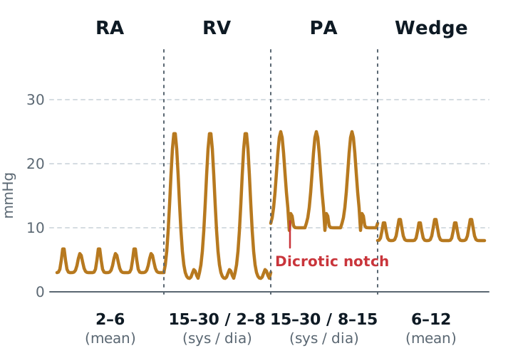
Read pressures at end-expiration, when intrathoracic pressure is closest to atmospheric, and average several breaths on a ventilator. Don’t leave the balloon inflated for more than about a minute. The wedge is a poor stand-in for left atrial pressure with severe mitral regurgitation, atrial fibrillation, or heavy lung disease.
Shock patterns
| Shock | CVP | PCWP | Cardiac output | SVR |
|---|---|---|---|---|
| Hypovolemic (blood or fluid loss) | Low | Low | Low | High |
| Cardiogenic (pump failure) | High | High | Low | High |
| Distributive (septic, anaphylactic) | Low or normal | Low or normal | Usually high (low late) | Low |
| Obstructive (tamponade, PE, tension pneumothorax) | High | Low or normal in PE; high in tamponade | Low | High |
Cardiogenic shock is usually defined by a CI of 2.2 or less and a PCWP above 15 mmHg. Septic shock is the classic low-SVR state. Static numbers like CVP and PCWP are weak predictors of fluid response, so look at the whole picture.
Cardiogenic versus noncardiogenic edema
A high wedge (classically above about 18 mmHg) points to cardiogenic pulmonary edema from a failing left heart. A normal or low wedge with bilateral infiltrates points to noncardiogenic edema such as ARDS. The Berlin ARDS definition no longer requires a wedge pressure, but the logic still helps: leaky capillaries, not high pressure.
Study example
A hypotensive patient has CVP 3, PCWP 5, CI 1.6, and a high SVR. The pressures are low and the output is low: hypovolemic shock, so the answer is volume. If the same patient had CVP 14, PCWP 24, and CI 1.7, the pump has failed, and volume would worsen the lung water. Check the lungs for crackles and the film for edema.
Related: physical assessment, hypoxemia, ECG rhythms, ARDS.
Sources
- Chang Y. Calculation of Pulmonary and Systemic Vascular Resistance. OpenAnesthesia. 2026.
- Yartsev A. Directly measured and derived PA catheter variables. Deranged Physiology.
- Suarez VR, Bukowski RK, Hankins GDV. Hemodynamic Monitoring of the Critically Ill Patient. Global Library of Women's Medicine.
- Merck Manual Professional Edition. Shock.
- Hendy A, Bubenek-Turconi SI. The diagnosis and hemodynamic monitoring of circulatory shock: current and future trends. J Crit Care Med. 2016.
- Hamzaoui O, et al. Hemodynamic monitoring in cardiogenic shock. 2023.