Heart failure and cardiogenic pulmonary edema
Tell cardiogenic edema from ARDS by signs, chest X-ray, BNP, and wedge pressure, and know what the RT does: upright, oxygen, CPAP or bilevel.
- PCWP, cardiogenic> about 18 mmHgOlder ARDS definitions needed under 18
- NT-proBNP: unlikely HF< 300 pg/mLAbove 900 (age 50–75) makes it likely
- First-line supportCPAP or bilevelCuts intubation in moderate to severe edema
Cardiogenic pulmonary edema is fluid in the lungs because the left side of the heart can’t move blood forward. Pressure backs up into the pulmonary veins and pushes fluid into the interstitium and alveoli. In ARDS the fluid comes from leaky vessels instead. The two look alike on the ventilator, but the treatment differs, so a good RT knows the clues.
What you find
History and exam: shortness of breath that starts or worsens when lying flat (orthopnea), waking up breathless (paroxysmal nocturnal dyspnea), fast breathing and low oxygen, and diffuse crackles. In severe cases, the patient coughs up frothy, sometimes blood-tinged sputum. Look for distended neck veins and listen for a third heart sound (S3); both point toward heart failure. Heart failure can also bring leg swelling.
Chest X-ray: an enlarged heart (cardiomegaly) is often the backdrop. As the pressure in the pulmonary veins rises, the lungs show these signs, roughly in this order:
- Cephalization: the upper-lobe vessels become more prominent as blood flow is redistributed upward.
- Peribronchial cuffing and Kerley B lines: short lines at the lung edges, perpendicular to the pleura, from fluid in the lymphatics. Small pleural effusions may appear.
- Bat-wing (butterfly) opacities: fluid fills the alveoli around the hila.
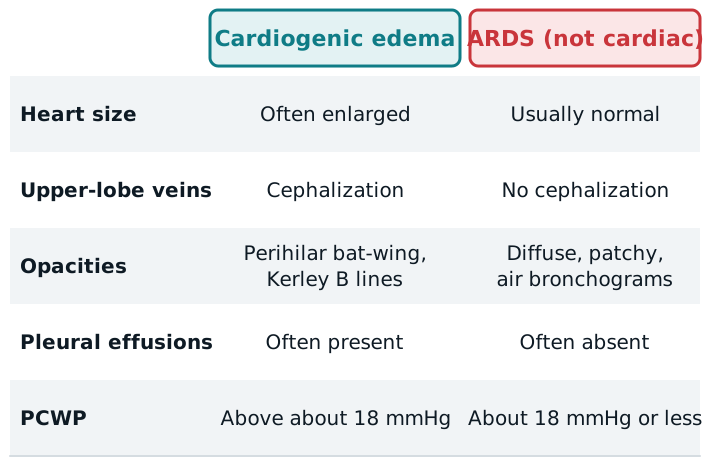
Cardiogenic or not?
In ARDS the heart is usually normal in size, vessels aren’t redistributed upward, effusions are often absent, and air bronchograms are common. The old consensus ARDS definition required a pulmonary artery wedge pressure (PCWP) under 18 mmHg; a higher value points to a cardiac cause. Pulmonary artery catheters are rarely used now, so the Berlin definition leans on a clinical look and echocardiography.
A blood test helps. Emergency guidance for acute heart failure uses NT-proBNP: under 300 pg/mL makes heart failure unlikely, and above 900 pg/mL (age 50 to 75) or 1,800 pg/mL (over 75) makes it likely. BNP works the same way with its own cutoffs. A high value supports heart failure but doesn’t prove it.
What the RT does
The team treats the cause with diuretics, nitroglycerin (if blood pressure allows), and other drugs. Your part is breathing support:
- Sit the patient upright, which eases orthopnea.
- Give oxygen, escalating from a mask to high-flow nasal oxygen or NIV.
- Use CPAP or bilevel NIV early. A 2026 review calls NIV the strongest-evidence option in moderate to severe edema: it reduces intubation and relieves dyspnea quickly. The positive pressure lowers preload and left ventricular afterload, which helps the drugs work.
- Choose between CPAP and bilevel by the patient’s CO2 and work of breathing; bilevel adds inspiratory pressure for a patient who is tiring or hypercapnic.
- High-flow nasal oxygen is an emerging alternative in milder cases, but the evidence is not as strong. See HFNC.
Tell the team early if the blood pressure drops, the patient grows tired, or the mask won’t seal.
Study example
A man with known heart failure arrives short of breath, sitting bolt upright because lying down is impossible. His SpO2 is 84% on room air, he has crackles to the mid-lungs, and the X-ray shows a large heart with bat-wing opacities. The team starts a diuretic and nitroglycerin. You start CPAP at a low pressure, titrate to the response, and reassess his work of breathing and SpO2 in the next few minutes.
Related: ARDS, COPD, ABG interpretation, pulse oximetry.
Sources
- Respiratory Support in Cardiogenic Pulmonary Edema: Clinical Insights from Cardiology and Intensive Care. J Cardiovasc Dev Dis. 2026.
- CAEP Acute Heart Failure Best Practices Checklist. CJEM. 2025.
- Chest radiography in adult critical care unit: a pictorial review. Indian J Radiol Imaging. 2019.
- Matthay MA, et al. Acute respiratory distress syndrome. Nat Rev Dis Primers. 2019.
- Murray and Nadel's Textbook of Respiratory Medicine: History and Physical Examination.