Pulmonary embolism: dead space and the ETCO2 clue
Why a clot in the lung raises dead space, what happens to ETCO2 and the PaCO2–ETCO2 gap, how PE is diagnosed, and how it is treated.
- Virchow's triad3 factorsStasis, hypercoagulability, endothelial injury
- ETCO2 in PEFallsPaCO2–ETCO2 gap widens (dead space)
- Imaging of choiceCT angiographyV/Q scan if CTA is not an option
- High-risk PEThrombolysisUnless contraindicated
A pulmonary embolism (PE) is a blood clot, usually from a leg or pelvic vein, that lodges in the pulmonary arteries. Blood can’t reach the alveoli beyond the clot, but the patient keeps ventilating them. That mismatch is why PE matters to an RT: oxygenation falls, and the CO2 numbers on the monitor change in a recognizable way.
Why clots form
Virchow’s triad lists the three factors behind thrombus formation: venous stasis (flow is slow, as after long immobility), hypercoagulability (inherited clotting disorders, cancer, pregnancy), and endothelial damage (injury, inflammation, trauma). Ask about recent surgery, immobility, cancer, and pregnancy.
What it does to the lungs
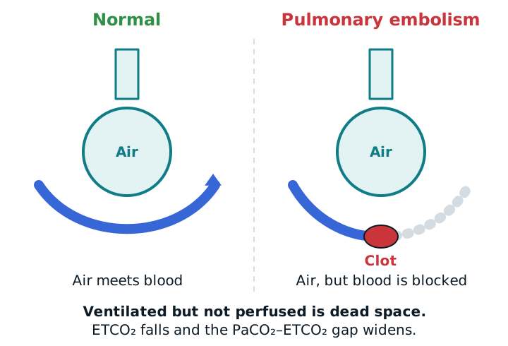
Alveoli beyond the clot are ventilated but not perfused. That is dead space: gas goes in and out without exchanging CO2. Dead space means the same minute ventilation clears less CO2, and the V/Q mismatch lowers oxygenation. A large clot also strains the right ventricle.
How it presents
Look for sudden shortness of breath, chest pain that hurts with breathing, a fast heart rate, and low SpO2. A large PE can cause fainting, low blood pressure, and shock. The ABG may show a low PaO2 with a low PaCO2 and respiratory alkalosis, plus a high A–a gradient from V/Q mismatch; one reported case had pH 7.46, PaCO2 33 mmHg, and an A–a gradient of about 95 mmHg. A normal ABG doesn’t exclude PE.
Capnography: the dead-space clue
When ventilated, unperfused alveoli add dead space, ETCO2 falls and the gap between PaCO2 and ETCO2 widens. In one 2026 study, low ETCO2 flagged high-severity PE and shock-level hemodynamics. In a ventilated patient, a sudden fall in ETCO2 without a circuit leak, together with falling SpO2 or blood pressure, should make you think of PE, though it doesn’t diagnose it on its own. See capnography.
Capnography can also mislead. In case reports of massive PE and cardiac arrest, the ETCO2 response was so weak that it looked as if the tube was not in the trachea, even though it was. In that setting, confirm tube position by direct view, not just by the waveform.
Diagnosis
- Pretest probability. Use a score such as Wells or Geneva to sort patients as low, intermediate, or high probability.
- D-dimer. It is highly sensitive, so a negative result, together with low or intermediate pretest probability (and the PERC rule), rules out PE without imaging. A positive D-dimer doesn’t confirm PE.
- CT pulmonary angiography (CTA) is the standard test. A V/Q scan is used in pregnancy, with contrast allergy or kidney disease, or when the patient can’t be moved. Echocardiography and leg ultrasound add information.
Treatment
The base of treatment is anticoagulation (unfractionated or low-molecular-weight heparin), started quickly. Systemic thrombolysis is considered for high-risk PE (hypotension or shock) if there are no contraindications. Catheter-directed therapy and embolectomy are options when thrombolysis is risky or fails. The 2026 AHA/ACC guideline replaced the older terms “massive” and “submassive” with categories A to E, so you will see both in textbooks. As the RT, you give oxygen, monitor SpO2 and ETCO2, and support the airway and ventilation as needed.
Study example
An intubated trauma patient on bed rest is on a steady ventilator setting. ETCO2 falls from 36 to 22 mmHg over a few minutes, SpO2 drops, and blood pressure falls. The circuit is intact and the tube is in place. An ABG shows a PaCO2 of 38 mmHg, so the PaCO2–ETCO2 gap is 16 mmHg and widening. That doesn’t prove a PE, because other causes of dead space and low perfusion exist, but it fits the picture. You tell the team what you see and why you are worried about ventilated, unperfused lung.
Related: capnography, ABG interpretation, ARDS, pulse oximetry.
Sources
- Rali P, et al. Diagnosis, Treatment and Follow Up of Acute Pulmonary Embolism: Consensus Practice from the PERT Consortium. Clin Appl Thromb Hemost. 2019.
- A PERT Consortium Appraisal of the 2026 AHA/ACC Multisociety Guideline for Acute Pulmonary Embolism. JACC Adv. 2026.
- Dead space in critical care: a practical approach with clinical scenarios. J Anesth Analg Crit Care. 2026.
- Low end-tidal carbon dioxide identifies high-severity pulmonary embolism and shock-level haemodynamics. ERJ Open Res. 2026.
- Genetic and biological determinants of pulmonary embolism: insights from Mendelian randomization studies. World J Exp Med. 2026.
- Potential pitfalls in diagnostic and therapeutic decision-making in pulmonary embolism-related obstructive shock. Eur Heart J Case Rep. 2026.