Endotracheal intubation: equipment and confirmation
Know the equipment, adult tube depth, how to confirm placement with waveform capnography, and how to spot a right mainstem tube.
- Depth at the teeth21–23 cmTypical adult; check on X-ray
- Tip on chest X-rayAbout 5 cm above carinaHead neutral; range about 2–6 cm
- Best confirmationWaveform capnographyContinuous ETCO2 trace
- Cuff pressure20–30 cm H2OMeasure with a manometer
Endotracheal (ET) intubation places a tube through the mouth or nose into the trachea to secure the airway and allow ventilation. The RT usually assists, then owns the checks afterward: is the tube in the trachea, at the right depth, with a safe cuff pressure?
When to intubate
Typical reasons are failure to oxygenate or ventilate that noninvasive support cannot fix, inability to protect the airway, and an expected decline. In a COPD exacerbation, the BTS guideline lists imminent respiratory arrest, severe distress, failed or contraindicated NIV, a pH that stays below 7.15 or worsens on NIV, and a GCS below 8. Preoxygenation with HFNC or NIV is strongly recommended in the 2026 ATS guideline to prevent desaturation during the attempt.
Equipment
| Item | Notes |
|---|---|
| Laryngoscope blade | A curved Macintosh blade tip goes into the vallecula and lifts the epiglottis indirectly. A straight Miller blade picks the epiglottis up directly. |
| Video laryngoscope | Camera at the blade tip. Guidelines suggest it for adult intubation, especially when the airway looks difficult. |
| Stylet | Stiffens the tube. Keep its tip 1–2 cm short of the tube end so the tip stays soft. |
| Bougie | A semirigid introducer passed under the epiglottis when the view is poor; the tube is then railroaded over it. |
| Tube | Most adults can take an inner diameter of 8 mm or more, which lowers resistance and eases suctioning and bronchoscopy. |
| Also ready | Suction, bag-valve-mask with oxygen, syringe for the cuff, capnography, cuff manometer, securement device. |
Depth
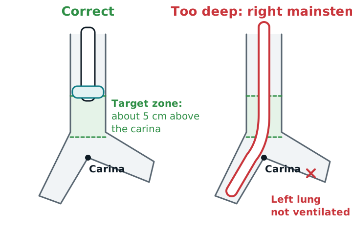
Typical adult depth is 21–23 cm at the teeth. A randomized trial found the average correct depth was 21 cm in women and 23 cm in men. But at those depths the tip was within 2.5 cm of the carina in about 1 in 5 patients, so the authors favored 20–21 cm in women and 22–23 cm in men. A chest X-ray should show the tip about 5 cm above the carina with the head neutral (a range of roughly 2–6 cm is accepted). Neck flexion moves the tube about 2 cm deeper and extension moves it about 2 cm out.
Confirming placement
- Look and listen as you bag: the tube passes between the cords, the chest rises equally, breath sounds are present on both sides, and there is no gurgling over the stomach.
- Waveform capnography is the standard of care and the gold standard. A continuous ETCO2 trace with normal-looking breaths confirms the tube is in the airway. Colorimetric detectors are acceptable but less complete. A flat trace means no ventilation, with one exception: in prolonged cardiac arrest or very low output, CO2 may be missing even with a correct tube.
- Chest X-ray confirms depth once the patient is stable.
Do not rely on auscultation alone. In one trial it missed more than a third of endobronchial intubations (sensitivity 65%), and first-year residents missed 55%.
Right mainstem signs
Breath sounds and chest rise are less on the left, peak pressures climb, SpO2 falls, and the tube reads deeper than before. Pull the tube back a little, recheck both sides, and confirm with an X-ray.
Cuff pressure
Keep the cuff at 20–30 cm H2O. Too low risks aspiration of secretions; too high reduces blood flow to the tracheal wall. Measure with a manometer. Palpating the pilot balloon is inaccurate.
Study example
A 5 ft 10 in man is intubated with an 8.0 tube at 23 cm at the teeth. The ETCO2 trace is steady, but the left chest rises less and SpO2 slips to 89%. The chest X-ray shows the tip in the right main bronchus. You deflate the cuff, withdraw the tube until both sides of the chest rise and sound equal, reinflate the cuff to 25 cm H2O, note the new depth at the teeth, and get a repeat X-ray.
See ventilator alarms and troubleshooting, capnography, chest X-ray basics, and airway suctioning.
Sources
- Merck Manual Professional Edition. Tracheal intubation.
- Frerk C, et al. Difficult Airway Society 2015 guidelines for management of unanticipated difficult intubation in adults. Br J Anaesth. 2015.
- Sitzwohl C, et al. Endobronchial intubation detected by insertion depth of endotracheal tube, bilateral auscultation, or observation of chest movements: randomised trial. BMJ. 2010.
- Mathew RP, et al. Chest radiographs of cardiac devices (Part 1): lines, tubes, non-cardiac medical devices and materials. SA J Radiol. 2019.
- Lohan R, Chawla A. Imaging of ICU patients. In: Thoracic Imaging. Springer; 2019.
- Goodfellow LT, et al. AARC Clinical Practice Guideline: Patient-ventilator assessment. Respir Care. 2024;69(8):1042-1054.